- a concentration of Na+ outside the cell that is some 10 times greater than that inside the cell
- a concentration of K+ inside the cell some 20 times greater than that outside the cell.
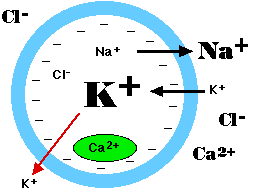
All cells (not just excitable cells) have a resting potential: an electrical charge across the plasma membrane, with the interior of the cell negative with respect to the exterior. The size of the resting potential varies, but in excitable cells runs about -70 millivolts (mv).
The resting potential arises from two activities:| Ionic relations in the cell. | |
The sodium/potassium ATPase produces
|
|
If depolarization at a spot on the cell reaches the threshold voltage, the reduced voltage now opens up hundreds of voltage-gated sodium channels in that portion of the plasma membrane. During the millisecond that the channels remain open, some 7000 Na+ rush into the cell. The sudden complete depolarization of the membrane opens up more of the voltage-gated sodium channels in adjacent portions of the membrane. In this way, a wave of depolarization sweeps along the cell. This is the action potential (In neurons, the action potential is also called the nerve impulse.)
The axons of many neurons are encased in a fatty sheath called the myelin sheath. It is the greatly expanded plasma membrane of an accessory cell called the Schwann cell. Where the sheath of one Schwann cell meets the next, the axon is unprotected. The voltage-gated sodium channels of myelinated neurons are confined to these spots (called nodes of Ranvier).
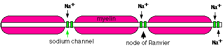
The inrush of sodium ions at one node creates just enough depolarization to reach the threshold of the next. In this way, the action potential jumps from one node to the next. This results in much faster propagation of the nerve impulse than is possible in nonmyelinated neurons.
A second stimulus applied to a neuron (or muscle fiber) less than 0.001 second after the first will not trigger another impulse. The membrane is depolarized and the neuron is in its refractory period. Not until the -70 mv polarity is reestablished will the neuron be ready to fire again.
Repolarization is first established by the facilitated diffusion of potassium ions out of the cell. Only when the neuron is finally rested are the sodium ions that came in at each impulse actively transported back out of the cell.
In some human neurons, the refractory period lasts only 0.001-0.002 seconds. This means that the neuron can transmit 500-1000 impulses per second.
Example: Gamma amino butyric acid (GABA). This neurotransmitter is found in the brain and inhibits nerve transmission by both mechanisms:
A single neuron, especially one in the central nervous system, may have thousands of other neurons synapsing on it. Some of these release activating (depolarizing) neurotransmitters; others release inhibitory (hyperpolarizing) neurotransmitters.
The receiving cell is able to integrate these signals. If (within a brief period) summation of the signals enables the cell to reach threshold, a nerve impulse is generated.
One might expect that depolarization at one point on the plasma membrane would generate an action potential irrespective of inhibitory signals elsewhere. However, this is avoided by having one portion of the plasma membrane (called the axon hillock) with| Welcome&Next Search |