Extraembryonic Membranes
 The embryos of reptiles, birds, and mammals produce 4 extraembryonic membranes, the
- amnion
- yolk sac
- chorion, and
- allantois
In birds and most reptiles, the embryo with its extraembryonic membranes develops within a shelled egg.
- The amnion protects the embryo in a sac filled with amniotic fluid.
- The yolk sac contains yolk - the sole source of food until hatching. Yolk is a mixture of proteins and lipoproteins.
- The chorion lines the inner surface of the shell (which is permeable to gases) and participates in the exchange of O2 and CO2 between the embryo and the outside air.
- The allantois stores metabolic wastes (chiefly uric acid) of the embryo and, as it grows larger, also participates in gas exchange.
With these four membranes, the developing embryo is able to carry on essential metabolism while sealed within the egg. Surrounded by amniotic fluid, the embryo is kept as moist as a fish embryo in a pond.
Although (most) mammals do not make a shelled egg, they do also enclose their embryo in an amnion. For this reason, the reptiles, birds, and mammals are collectively referred to as the amniota.
Mammals fall into three groups that differ in the way they use the amniotic egg.
- Monotremes
These primitive mammals produce a shelled egg like their reptilian ancestors. Only three species exist today: two species of spiny anteater (echidna) and the duckbill platypus. [View (48K)]
- Marsupials
Marsupials do not produce a shelled egg. The egg, which is poorly supplied with yolk, is retained for a time within the reproductive tract of the mother. The embryo penetrates the wall of the uterus. The yolk sac provides a rudimentary connection to the mother's blood supply from which it receives food, oxygen, and other essentials. However, this interface between the tissues of the uterus and the extraembryonic membranes never becomes elaborately developed, and the young are born in a very immature state.
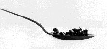
The photo (courtesy of Dr. Carl G. Hartman) shows 18 newborn baby opossums fitting easily into a teaspoon.
Despite their tiny size, they are able to crawl into a pouch on the mother's abdomen, attach themselves to nipples, and drink milk from her mammary glands.
Marsupials are still abundant in Australia, but only the opossum is found in North America.
- Placental mammals
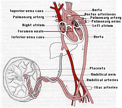
In placental mammals, the extraembryonic membranes form a placenta and umbilical cord, which connect the embryo to the mother's uterus in a more elaborate and efficient way. The blood supply of the developing fetus is continuous with that of the placenta. [View below] The placenta extracts food and oxygen from the uterus. Carbon dioxide and other wastes (e.g., urea) are transferred to the mother for disposal by her excretory organs.
Embryonic development begins while the fertilized egg is still within the fallopian tube. The developing embryo travels down the tube, reaching the uterus in three or four days. As a result of repeated mitotic divisions and some migration of cells, a hollow ball of cells is formed called the blastocyst. Approximately one week after fertilization, the blastocyst embeds itself in the thickened wall of the uterus, a process called implantation, and pregnancy is established.
The blastocyst produces two major divisions of cells:
- Three or four blastocyst cells develop into the inner cell mass, which will become the fetus and, ultimately, the baby.
- The remaining 100 or so cells form the trophoblast, which will produce the four extraembryonic membranes. These will play vital roles during development but will be discarded at the time of birth.
The extraembryonic membranes form the amnion, placenta and umbilical cord.
The placenta grows tightly fused to the wall of the uterus. Its blood vessels, supplied by the fetal heart, are literally bathed in the mother's blood. Although there is normally no mixing of the two blood supplies, the placenta does facilitate the transfer of a variety of materials between the fetus and the mother.
- receiving food
- receiving oxygen and discharging carbon dioxide
- discharging urea and other wastes
- receiving antibodies (chiefly of the IgG class). These remain for weeks after birth, protecting the baby from the diseases to which the mother is immune.
But the placenta is not simply a transfer device. Using raw materials from the mother's blood, it synthesizes large quantities of proteins and also some hormones.
The metabolic activity of the placenta is almost as great as that of the fetus itself.
The umbilical cord connects the fetus to the placenta. It receives deoxygenated blood from the iliac arteries of the fetus and returns oxygenated blood to the liver and on to the inferior vena cava.
Because its lungs are not functioning, circulation in the fetus differs dramatically from that of the baby after birth. While within the uterus, blood pumped by the right ventricle bypasses the lungs by flowing through the foramen ovale and the ductus arteriosus.
Although the blood in the placenta is in close contact with the mother's blood in the uterus, intermingling of their blood does not normally occur. However, some of the blood cells of the fetus usually do get into the mother's circulation - where they have been know to survive for decades. This raises the possibility of doing prenatal diagnosis of genetic disorders by sampling the mother's blood rather than having to rely on the more invasive procedures of amniocentesis and chorionic villus sampling (CVS).
Far rarer is the leakage of mother's blood cells into the fetus. However, it does occur. A few pregnant women with leukemia or lymphoma have transferred the malignancy to their fetus. Some babies have also acquired melanoma from the transplacental passage of these highly-malignant cells from their mother.
The placenta is an allograft
One of the greatest unsolved mysteries in immunology is how the placenta survives for 9 months without being rejected by the mother's immune system. Every cell of the placenta carries the father's genome (a haploid set of his chromosomes); including one of his #6 chromosomes where the genes for the major histocompatibility antigens are located.
| One partial exception: none of the genes on the father's X chromosome are expressed. While X-chromosome inactivation is random in the cells of the fetus, it is NOT random in the cells of the trophoblast. In every cell of the trophoblast - and its descendants - it is the paternal X chromosome that is inactivated. [Discussion of X-chromosome inactivation.] But this does not solve our problem because the genes for all the major histocompatibility antigens are located on chromosome 6, which is not inactivated. |
| Discussion of the human major histocompatibility complex (MHC) |
Thus the placenta is immunologically as foreign to the mother as a kidney transplant would be.
Yet it thrives.
Despite a half-century of research, the mechanism for this immunologically privileged status remains uncertain. But one thing is clear:
The mother is not intrinsically tolerant of the father's antigens.
Some evidence:
- She will promptly reject a skin transplant from the father.
- She develops antibodies against his histocompatibility antigens expressed by the fetus. In fact, women who have borne several children by the same father are often excellent sources of anti-HLA serum for use in tissue typing.
So what accounts for the phenomenon? Some possibilities:
- The placenta does not express class II histocompatibility antigens.
- The class I histocompatibility antigens that it does express are only weakly immunogenic.
- A recent report shows that in normal mice, the cells of the placenta degrade the amino acid tryptophan. Tryptophan is essential for T-cell function. When mice were treated with an inhibitor of the Trp-degrading enzyme, their fetuses were promptly aborted by the action of the mother's lymphocytes. (D. H. Munn, et. al., Science, 281: 1191, 21 Aug 1998.)
17 August 1999