| Discussion of antigen presentation to B cells and T cells |
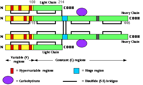
This image represents the polypeptide chain structure of a molecule of IgG. The numbers indicate the number of amino acids (counting from the amino terminal ("N"). In the actual molecule, the chains are folded to that each cysteine is brought close to the partner with which it forms a disulfide (S-S) bridge.
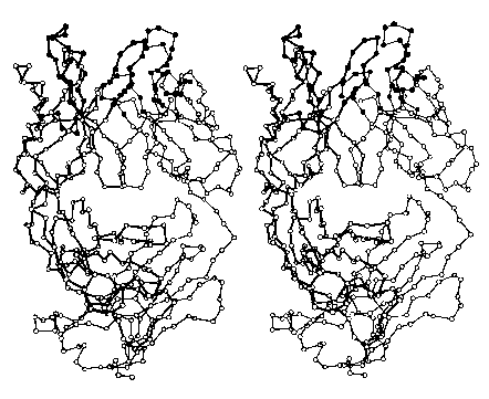
The images below (courtesy of Dr. D. R. Davies) represent the folded (tertiary) structure of an entire L chain (right side with thin connecting lines) and the V region plus the first third of the C region of a heavy chain (left side; darker lines). Each circle represents the location in 3D space of an alpha carbon. The filled circles at the top are amino acids in the hypervariable regions; they form the site that binds the antigen.
Do try to fuse these two images into a stereoscopic (3D) view. I find that it works best when my eyes are about 18" from the screen and I try to relax so that my eyes are directed at a point behind the screen.
Antibody molecules have two functions to perform:
So, V regions finger the culprit; the C regions take action.
Why 5 kinds of heavy chains? To provide for different effector functions.
| Class | H chain | L chain | Subunits | mg/ml | Notes |
|---|---|---|---|---|---|
| IgG | gamma | kappa or lambda | H2L2 | 13 | transferred across placenta |
| IgM | mu | kappa or lambda | (H2L2)5 | 0.5-2.5 | first antibodies to appear after immunization |
| IgA | alpha | kappa or lambda | (H2L2)2 | 0.5-3 | much higher concentrations in secretions |
| IgD | delta | kappa or lambda | H2L2 | 0.03 | function uncertain |
| IgE | epsilon | kappa or lambda | H2L2 | 0.0003 | binds to basophils and mast cells sensitizing them for certain allergic reactions |
The antigen receptor on most T cells is made up of two transmembrane polypeptides designated alpha and beta (thus forming a heterodimer).
Like antibodiesA small percentage of the T cells in the blood use a TCR consisting of a heterodimer of two other types of transmembrane polypeptides: gamma and delta. The function of this subset of T cells is still a mystery.
All the antibody Heavy chains, lambda L chains, kappa L chains, T cell alpha, beta, gamma, and delta chains are encoded by genes that are assembled from separate gene segments during the differentiation of the cell. The result is a single gene that is transcribed into a mRNA to be translated into one chain of the receptor. The number of gene segments from which the variable regions are constructed is sufficiently large that both B cells and T cells can generate as many as 1010 different antigen-binding sites. There is probably no epitope that could exist for which BCRs and TCRs able to bind it are not be built.
| The mechanisms by which B cells and T cells generate receptors of such extraordinary diversity. |
| Welcome&Next Search |