Animal Tissues
The development of a fertilized egg into a newborn child requires an average of 41 rounds of mitosis (241 = 2.2 x 10¹²).
During this period, the cells produced by mitosis enter different pathways of differentiation; some becoming blood cells, some muscle cells, and so on.
There are more than 100 visibly-distinguishable kinds of differentiated cells in the vertebrate animal. These are organized into tissues; the tissues into organs. Groups of organs make up the various systems - digestive, excretory, etc. - of the body.
The actual number of differentiated cell types is surely much larger than 100.
- All lymphocytes, for example, look alike but actually represent a variety of different functional types, e.g., B cells, T cells of various subsets.
- The neurons of the central nervous system must exist in hundreds of different functional types, each representing the result of a particular pathway of differentiation.
|
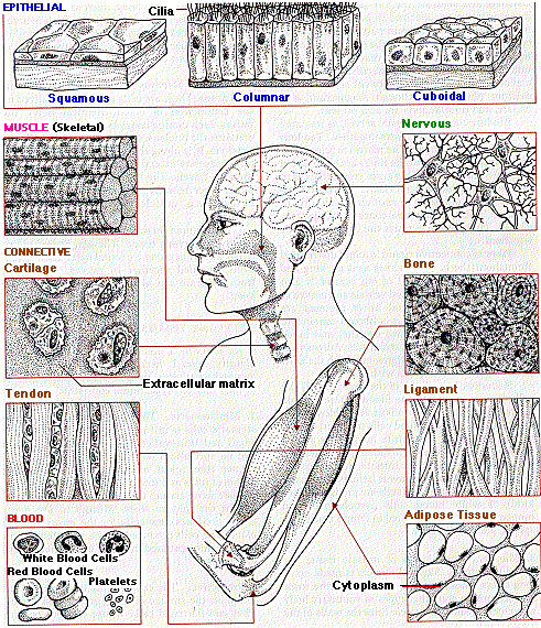
This page will give a brief introduction to the major types of animal tissues. The links along the left side of the figure will take you directly to the individual paragraphs indicated.
Epithelial tissue is made of closely-packed cells arranged in flat sheets. Epithelia line the various cavities and tubes of the body. They also form the surface of the skin.
The apical surface of epithelial cells is exposed to the "external environment", lumen of the organ or the air.
The basolateral surface is exposed to the internal environment (ECF). The entire sheet of epithelial cells is attached to a layer of extracellular matrix that is called the basement membrane (but it is not a membrane in the biological sense) or, better, the basal laminar.
The function of epithelia always reflects the fact that they are boundaries between masses of cells and a cavity or space. Some examples:
- The epithelium of the skin protects the underlying tissues from
- mechanical damage
- ultraviolet light
- dehydration
- invasion by bacteria
- The columnar epithelium of the intestine
- secretes digestive enzymes into the intestine
- absorbs the products of digestion from it.
- An epithelium also lines our air passages and lung cavities. It secretes mucus which keeps it from drying out and to trap inhaled dust particles. Most of its cells have cilia on their apical surface that propel the mucus with its load of foreign matter back up to the throat.
Three kinds of muscle are found in vertebrates:
- Skeletal muscle is made of long fibers whose contraction provides the force of locomotion and other voluntary body movements.
- Smooth muscle lines the walls of the hollow structures of the body, such as the intestine, urinary bladder, uterus, and blood vessels. Its contraction, which is involuntary, reduces the size of these hollow organs.
- The heart is made of cardiac muscle.
The cells of connective tissue are embedded in a great amount of extracellular material. This matrix is secreted by the cells. It consists of protein fibers embedded in an amorphous mixture of protein-polysaccharide ("proteoglycan") molecules.
Supporting connective tissue
Gives strength, support, and protection to the soft parts of the body.
- cartilage. Example: the outer ear
- bone. The matrix of bone contains collagen fibers and mineral deposits. The most abundant mineral is calcium phosphate, although magnesium, carbonate, and fluoride ions are also present.
Binding connective tissue
It binds body parts together.
- Tendons connect muscle to bone. [View] The matrix is principally collagen, and the fibers are all oriented parallel to each other. Tendons are strong but not elastic.
- Ligaments attach one bone to another. They contain both collagen and also the protein elastin. Elastin permits ligaments to be stretched.
Fibrous connective tissue
It is distributed throughout the body. It serves as a packing and binding material for most of our organs. Collagen, elastin, and other proteins are found in the matrix.
Fascia is fibrous connective tissue that binds muscle together and binds the skin to the underlying structures.
Adipose tissue is fibrous connective tissue in which the cells have become almost filled with oil. The cells of adipose tissue, called adipocytes, secrete the hormone leptin.
All forms of connective tissue are derived from cells called fibroblasts, which secrete the extracellular matrix.
Nerve tissue is composed principally of nerve cells called neurons. They are specialized for the conduction of nerve impulses. A typical neuron consists of
- a cell body which contains the nucleus
- a number of short fibers - dendrites - extending from the cell body
- a single long fiber, the axon.
The nerve impulse is conducted along the axon. The tips of axons meet:
- other neurons at junctions called synapses
- muscles
- glands
The bone marrow is the source of all the cells of the blood. These include:
- red blood cells (RBCs or erythrocytes)
- five kinds of white blood cells (WBCs or leukocytes)
- platelets or thrombocytes
3 August 1999