Organizing the Embryo: The Central Nervous System
In the embryonic development of a zygote, gradients of mRNAs and proteins, deposited in the egg by the mother as she formed it, give rise to cells of diverse fates despite their identical genomes.
But is the embryo fully patterned in the fertilized egg? It is difficult to imagine that the relatively simple gradients in the egg could account for all the complex migration and differentiation of cells during embryonic development. And, in fact, the answer is no. However, once these gradients have sent certain cells along a particular path of gene expression, the stage is set for those cells to begin influencing nearby cells to become increasingly diversified.
In other words,
- cell-intrinsic signals (established between a nucleus and the particular cytoplasmic environment that cleavage has placed it in) lay the foundation for
- cell-cell interactions to further guide the cells of the embryo to assume their proper position in the embryo and to differentiate into their final specialized form and function.
The Organizer
In 1924, the German embryologists Hans Spemann and Hilde Mangold performed an experiment that
- demonstrated that the pattern of development of cells is influenced by the activities of other cells and
- stimulated a search, which continues to this day, for the signals at work.
Spemann and Mangold knew that the cells that develop in the region of the gray crescent migrate into the embryo during gastrulation and form the notochord (the future backbone; made of mesoderm).
They cut out a piece of tissue from the gray crescent region of one newt gastrula and transplanted it into the ventral side of a second newt gastrula. To make it easier to follow the fate of the transplant, they used the embryo of one variety of newt as the donor and a second variety as the recipient.
(You may need to open your browser wider.)
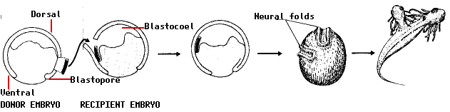
The remarkable results: - the transplanted tissue developed into a second notochord
- neural folds developed above the extra notochord
- these went on to form a second central nervous system (portions of brain and spinal cord) and eventually
- a two-headed tadpole.
But the most remarkable finding of all was that the neural folds were built from recipient cells, not donor cells. In other words, the transplant had altered the fate of the overlying cells (which normally would have ended up forming skin [epidermis] on the side of the animal) so that they produced a second head instead!
Spemann and Mangold used the term induction for the ability of one group of cells to influence the fate of another. And because of the remarkable inductive power of the gray crescent cells, they called this region the organizer.
Over the next three quarters of a century, vigorous searches have been made to identify the molecules liberated by the organizer that induce overlying cells to become nerve tissue. One candidate after another has been put forward and then found not to be responsible. Part of the problem has been that not until just recently has it become clear that the organizer
- does NOT induce the central nervous system but, instead,
- it prevents signals originating from the ventral side of the blastula from inducing skin (epidermis) there.
This is how it works:
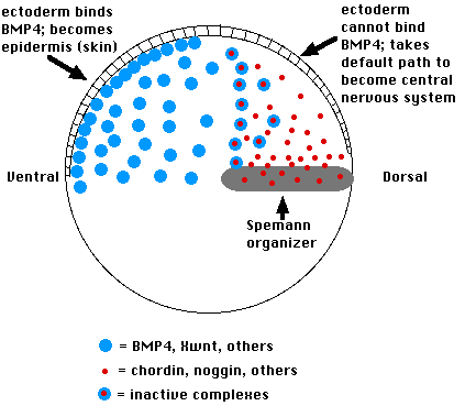
- Cells on the ventral side of the blastula secrete a variety of proteins such as bone morphogenetic protein-4 (BMP-4)
- These induce the ectoderm above to become epidermis.
- If their action is blocked, the ectodermal cells are allowed to follow their default pathway, which is to become nerve tissue of the brain and spinal cord.
- The Spemann organizer blocks the action of BMP-4 by secreting molecules of the proteins
- Both of these physically bind to BMP-4 molecules in the extracellular space and thus prevent BMP-4 from binding to receptors on the surface of the overlying ectoderm cells.
- This allows the ectodermal cells to follow their intrinsic path to forming neural folds and, eventually, the brain and spinal cord.
Patterning the central nervous system in Drosophila
Remarkably, it turns out that proteins similar in structure to the bone morphogenetic proteins and also to chordin are found in Drosophila.
- The role of BMP-4 is taken by a related protein encoded by the decapentaplegic gene (dpp).
- The role of chordin is taken by a related protein called sog encoded by the gene called short gastrulation.
In fact, these proteins and their mRNAs are completely interchangeable!
- an injection of the mRNAs for BMP-4 or chordin into the blastoderm of the Drosophila embryo can replace the function of dpp and sog respectively, and
- conversely, injections of mRNA for dpp or sog into the Xenopus embryo mimics the functions of BMP-4 and chordin respectively.
A selection of antagonistic pairs of proteins that guide the patterning of the embryo.
| Xenopus | Bone Morphogenetic Protein-4 (BMP-4) | blocked by chordin |
|---|
| and also by noggin |
| Drosophila | Decapentaplegic (DPP) | blocked by short gastrulation (SOG) |
|---|
| and also by a noggin homolog? |
| Mammal | several BMPs | blocked by ? |
|---|
Dorsal vs Ventral Nerve Cords
Although their actions are similar, the distribution of these proteins in Drosophila differs from that in Xenopus (as well as in mammals and other vertebrates).
In Drosophila,
- DPP is produced in the dorsal region of the embryo and
- SOG is produced in the ventral region.
However, their actions on overlying cells are the same as in Xenopus; that is,
the sog protein prevents the dpp protein from blocking the formation of the central nervous system.
The result in Drosophila is that its central nervous system forms on the ventral side of the embryo, not on the dorsal! And, you may remember that one of the distinguishing traits of all arthropods (insects, crustaceans, arachnids) as well as many other invertebrates, such as the annelid worms, is a ventral nerve cord. Vertebrates have a dorsal (spinal) nerve cord.
Patterning in Mammals
The establishment of the dorsal central nervous system probably occurs in mammals by mechanisms similar to those in Xenopus. Close relatives (homologs) of the Xenopus molecules occur in mammals. In fact, the BMPs were discovered in mammalian tissue before they were found in Xenopus (and have many important functions throughout the life of the animal). Similarly, the Wnt molecules (see below) found in Xenopus were first identified in mammals (as the product of oncogenes).
The patterning of the embryo employs a number of redundant, or at least overlapping, mechanisms. (In fact, genetic redundancy seems to be a key feature of life. It is the reason that knockout mice are so often able to function without certain genes).
The table below gives another set of antagonistic pairs of molecules:
- those in the left column block formation of nerve tissue;
- those in the right column bind to those in the left and thus permit the formation of nerve tissue.
Another set of antagonistic pairs of proteins that guide the patterning of the embryo.
| Xenopus | Xwnt-8 | Xfrzb-1 |
|---|
| Drosophila | wingless (wg) | frizzled |
|---|
| Mammal | various Wnt oncoproteins | frzb-1 ("frizbee") |
|---|
Once again, the molecules in one column are close relatives, even though many millions of years have passed since the ancestors of these animals took separate evolutionary paths!
We're half-way done!
Xenopus development (and probably that of animals in general) passes through three rather different (although often overlapping) phases:
- establishing the main axes (dorsal-ventral; anterior-posterior; left-right). This is done by gradients of mRNAs and proteins encoded by the mother's genes and placed in the egg by her.
- establishing the main body parts such as the notochord and central nervous system in vertebrates (discussed here and also described in Frog Embryology) and the segments in Drosophila
These are run by genes of the zygote itself.
- filling in the details; that is, building the various organs of the animal. (Our examples will include the wings, legs, and eyes of Drosophila.)
23 June 1999