The three-dimensional structure of the entire polypeptide chain.
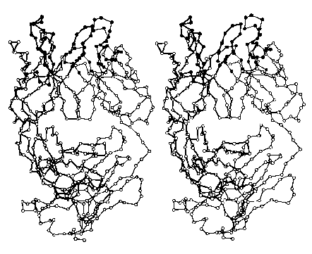
The images (courtesy of Dr. D. R. Davies) represent the tertiary structure of the antigen-binding portion of an antibody molecule. Each circle represents an alpha carbon in one of the two polypeptide chains that make up this protein. (The filled circles at the top are amino acids that bind to the antigen.) Most of the secondary structure of this protein consists of beta conformation, which is particularly easy to see on the right side of the image.
Do try to fuse these two images into a stereoscopic (3D) view. I find that it works best when my eyes are about 18" from the screen and I try to relax so that my eyes are directed at a point behind the screen.
Where the entire protein or parts of a protein are exposed to water (e.g., in blood or the cytosol), hydrophilic R groups are found at the surface; hydrophobic R groups are buried in the interior.
| Primary structure |
| Secondary structure |
| Quaternary structure |
| Welcome&Next Search |