Allergies
Immunologists, as well as the general public, use the term allergy in several different ways.
I shall define it thus:
An allergy is a harmful immune response elicited by an antigen that is not itself intrinsically harmful.
Examples:
- The windblown pollen released by orchard grass has no effect on me but produces a violent attack of hay fever (known to physicians as allergic rhinitis) in my wife.
- She, on the other hand, can safely handle the leaves of poison ivy while if I do so, I break out in a massive skin rash a day or two later.
Antigens that trigger allergies are often called allergens.
Four different immune mechanisms can result in allergic responses.
1. Immediate Hypersensitivities.
These occur quickly after exposure to the allergen. They are usually mediated by antibodies of the IgE class.
Examples:
2. Antibody-Mediated Cytotoxicity
Cell damage caused by antibodies directed against cell surface antigens. Hence a form of autoimmunity.
Examples:
- Hemolytic disease of the newborn (Rh disease).
- Myasthenia gravis (MG)
3. Immune Complex Disorders
Damage caused by the deposit in the tissues of complexes of antigen and their antibodies.
Examples:
- Serum sickness
- Systemic lupus erythematosus (SLE)
4. Cell-Mediated Hypersensitivities
These reactions are mediated by CD4+ T cells.
Examples:
- The rash produced following exposure to poison ivy.
Because it takes a day or two for the T cells to mobilize following exposure to the antigen, these responses are called delayed-type hypersensitivities (DTH). Those, like poison ivy, that are caused by skin contact with the antigen are also known as contact sensitivities or contact dermatitis.
- certain autoimmune diseases, including
- Insulin-dependent diabetes mellitus (IDDM)
- Multiple sclerosis (MS)
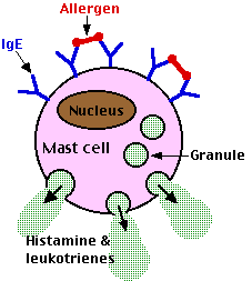
The constant region of IgE antibodies (shown in blue) has a binding site for a receptor present on the surface of basophils and their tissue-equivalent the mast cell. These cell-bound antibodies have no effect until and unless they encounter allergens (shown in red) with epitopes that can bind to their antigen-binding sites.
When this occurs, the mast cells to which they are attached explosively discharge their granules by exocytosis. The granules contain a variety of active agents including histamine and leukotrienes. Release of these substances into the surrounding tissue causes local anaphylaxis: swelling, redness, and itching. In effect, each IgE-sensitized mast cell is a tiny bomb that can be exploded by a particular antigen.
The most common types of local anaphylaxis are:
- allergic rhinitis (hay fever) in which airborne allergens react with IgE-sensitized mast cells in the nasal mucosa and the tissues around the eyes;
- bronchial asthma in which the allergen reaches the lungs either by inhalation or in the blood;
- hives (physicians call it urticaria) where the allergen usually enters the body in food.
Leukotrienes are far more potent than histamine in mediating these reactions.
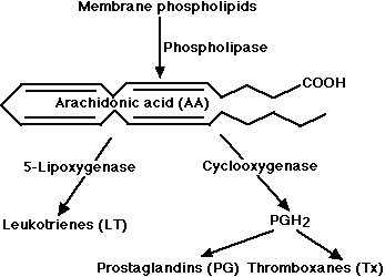
Leukotrienes and prostaglandins are derivatives of arachidonic acid (AA) an unsaturated fatty acid produced from membrane phospholipids. The principal pathways of arachidonic acid metabolism are
- the 5-lipoxygenase pathway, which produces a collection of leukotrienes (LT) and
- the cyclooxygenase pathway, which yields a number of prostaglandins (PG) and thromboxanes (Tx).
All three are synthesized by monocytes and macrophages. Mast cells and basophils generate a mixture of leukotrienes. The products of both pathways act in concert to cause inflammation with prostaglandins producing fever and pain. Aspirin, ibuprofen, and certain other nonsteroidal anti-inflammatory drugs (NSAIDs) achieve their effects (fever and pain reduction) by blocking the activity of cyclooxygenase. |
 |
Some people respond to environmental antigens (e.g., pollen grains, mold spores) with an unusually vigorous production of IgE antibodies. Why this is so is unclear; heredity certainly plays a role. In any case, the immune system of these people is tilted toward the production of T helper cells of the Th2 subtype. These release interleukin 4 (IL-4) and interleukin 13 (IL-13) on the B cells that they "help". These lymphokines promote class switching in the B cell causing it to synthesize IgE antibodies.
An inherited predisposition to making IgE antibodies is called atopy. Atopic people are apt to have higher levels of circulating IgE (up to 12 µg/ml) than is found usually (about 0.3 µg/ml). Whereas only 20-50% of the receptors on mast cells are normally occupied by IgE, all the receptors may be occupied in atopic individuals.
Skin Testing
When the problem allergen is not obvious, it can often be identified by skin testing. A panel of suspected allergens is injected into separate sites in the skin and each site is observed for the development of a "wheal and flare" reaction. The
- wheal is a sharply delineated soft swelling surrounded by the
- flare - a reddened area.
Both are caused by the release of leukotrienes at the site, which increase the flow of blood to the site making it swollen and red.
A positive skin test occurs within minutes or even seconds (in contrast to patch testing for DTH responses described below). In some patients, a response can be elicited by as little as 0.1 ng of allergen.
Some allergens can precipitate such a massive IgE-mediated response that a life-threatening collapse of the circulatory and respiratory systems may occur.
Frequent causes:
- insect (e.g., bee) stings
- many drugs (e.g., penicillin)
- a wide variety of foods (shellfish and nuts are common offenders; in fact, some school systems in the U. S. now ban peanuts and peanut-butter sandwiches when they have a student at risk of systemic anaphylaxis from exposure to peanuts.)
Treatment of systemic anaphylaxis centers on the quick administration of adrenaline, antihistamines, and - if shock has occurred - intravenous fluid replacement.
In these disorders, the person produces antibodies directed against antigens present on the surface of his or her own cells. Thus these qualify as autoimmune disorders.
Some examples:
Binding of antibodies to the surface of the cell can result in:
- phagocytosis of the cell
- lysis of the cell
- damage to molecules on the cell surface (e.g., myasthenia gravis)
- activation of cell-surface receptors (e.g., thyrotoxicosis)
Rh antigens are expressed at the surface of red blood cells.
During pregnancy, there is often a tiny leakage of the baby's red blood cells into the mother's circulation. If the baby is Rh-positive (having inherited the trait from its father) and the mother Rh-negative, these red cells will cause her to develop antibodies against the Rh antigen. The antibodies, usually of the IgG class, may not develop fast enough to cause problems for that child, but can cross the placenta and attack the red cells of a subsequent Rh+ fetus. This destroys the red cells producing anemia and jaundice. The disease may be so severe as to kill the fetus or even the newborn infant.
Although certain other red cell antigens (in addition to Rh) sometimes cause problems for a fetus, an ABO incompatibility does not. Why is an Rh incompatibility so dangerous when ABO incompatibility is not?
It turns out that most anti-A or anti-B antibodies are of the IgM class and these do not cross the placenta. In fact, an Rh-/type O mother carrying an Rh+/type A, B, or AB fetus is resistant to sensitization to the Rh antigen. Presumably her anti-A and anti-B antibodies destroy any fetal cells that enter her blood before they can elicit anti-Rh antibodies in her.
This phenomenon has led to an extremely effective preventive measure to avoid Rh sensitization. Shortly after each birth of an Rh+ baby, the mother is given an injection of anti-Rh antibodies. The preparation is called Rh immune globulin (RhIG) or Rhogam. These passively acquired antibodies destroy any fetal cells that got into her circulation before they can elicit an active immune response in her.
Rh immune globulin came into common use in the United States in 1968, and within a decade the incidence of Rh hemolytic disease became very low.
Some people synthesize antibodies against their own red blood cells, and these may lyze the cells producing anemia. Infections, cancer, or an autoimmune disease like systemic lupus erythematosus (SLE) are often involved. Many drugs (e.g. penicillin, quinidine) can also trigger the disorder. In these cases, stopping the drug usually brings about a quick cure.
This is an autoimmune disorder in which the patient develops antibodies against his or her own platelets (thrombocytes). The life span of the platelets may be reduced from the normal of 8 days to as little as 1 hour, and platelet counts may drop from a normal of 250,000/µl to 20,000/µl. This greatly interferes with normal clotting, causing
- external bleeding (e.g., from the nose) and
- internal bleeding into the skin causing purple patches (called purpura).
Often no cause of the disorder can be found (the physicians call it "idiopathic"). Some cases are triggered by prescription drugs like aspirin, digitoxin, and sulfa drugs. These cases can be cured by stopping the drug. The idiopathic cases can sometimes be helped by giving corticosteroids and/or removing the patient's spleen.
The hallmark of this autoimmune disorder is weakness of the skeletal muscles, especially those in the upper part of the body. It is caused by antibodies that attack the acetylcholine (ACh) receptors at the subsynaptic membrane of neuromuscular junctions. As the number of receptors declines, the ACh released with the arrival of a volley of nerve impulses is inadequate to generate end-plate potentials (EPPs) of the normal size. After repeated stimulation, the EPPs fail to reach the threshold needed to generate an action potential and the muscle stops responding.
The signs and symptoms of myasthenia gravis can be quickly - but only temporarily - relieved by injecting a drug that inhibits the action of cholinesterase. This prolongs the action of ACh at the neuromuscular junction. The immunosuppressant action of corticosteroids, like prednisone, can provide longterm improvement for patients.
The exclusive role of antibodies (of the IgG class) in this disorder is demonstrated by the presence of the disease in the newborn babies of mothers with the disorder. As these antibodies, which the fetus had received from the mother's circulation, disappear (in 1 -2 weeks), so do all signs of the disease.
In this disorder, the patient has antibodies that bind to the TSH receptors on the thyroxine-secreting cells of the thyroid. These antibodies mimic the action of TSH itself (thus they behave as a TSH agonist) and trigger secretion of thyroxine (T4) and T3 by the thyroid gland.
| Links to a discussion of the
|
The role of antibodies (of the IgG class) in this disorder is demonstrated by the presence of the disease in the newborn babies of mothers with the disorder. As these antibodies, which the fetus had received from the mother's circulation, disappear (in 1 -2 weeks), so do all signs of the disease.
While binding of antibody to antigen is often a helpful - even life-saving - response, in some circumstances it causes pathological changes.
In passive immunization, an antiserum containing needed antibodies is injected into the patient. At one time, these antisera were prepared by immunizing horses or sheep. While they did their intended work,
(usually to provide immediate protection to a person exposed to
they also often later lead to a syndrome called serum sickness. The patient developed
- fever
- hives
- arthritis and
- protein in the urine.
After a week or two, the symptoms would disappear spontaneously.
Serum sickness is caused by the many extraneous proteins present in the antiserum. Being foreign to the recipient, an active immunity develops against these proteins. The resulting antibodies bind to them forming immune complexes. These are carried by the blood and deposited in the walls of blood vessels as well as in the glomeruli of the kidneys.
Antigen-antibody complexes bind to a system of serum proteins collectively known as complement. The complex of antigen-antibody-complement
attracts basophils and mast cells and causes them to release their histamine and leukotrienes producing inflammation.
Thanks to nearly universal active immunization against both tetanus and diphtheria, serum sickness is now quite rare. However, kidney damage (called glomerulonephritis) produced by deposits of immune complexes is found in other ailments.
Persistent infections
Some infectious agents live in the body for long periods
Examples:
- the protozoans that cause malaria
- the worms that cause schistosomiasis and filariasis
- the virus that causes hepatitis B.
In these cases, the continued presence of the pathogen provides a renewable source of antigen to combine with antibodies synthesized by the host resulting in deposits of immune complexes.
Humans with SLE develop (for unknown reasons) antibodies against a wide variety of self components:
- their own DNA and RNA
- red blood cells
- platelets
- ribosomes
- even their own IgG molecules. (These "anti-antibodies" are called rheumatoid factors. They are also found in people with rheumatoid arthritis (hence the name) and, for a time, in people with mononucleosis.)
In all these cases of autoimmunity, immune complexes form and are deposited in the skin, joints, and kidneys where they initiate inflammation.
Repeated exposure to airborne organic particles, like mold spores, can elicit formation of antibodies. When these interact with inhaled antigen, inflammation of the alveoli occurs. The sufferer develops a cough, fever, and difficulty in breathing. Once removed from the source of antigen, the attack subsides within a few days.
Farmers exposed to moldy hay often develop this problem (technically known as extrinsic allergic alveolitis). Sugarcane workers, cheese makers, mushroom growers, pigeon fanciers, and a number of other occupational or hobby groups are apt to develop allergic alveolitis from exposure to the spores and dusts associated with their activities.
Cell-mediated hypersensitivities can occur with extrinsic antigens or with internal ("self") antigens.
The most common example of cell-mediated hypersensitivity to external antigens is the contact dermatitis caused in some people when their skin is exposed to a chemical to which they are allergic.
Some examples:
- the catechols found in poison ivy, poison oak, and poison sumac
- nickel (often used in jewelry)
- some dyes
- certain organic chemicals used in industry
In every case, these simple chemicals probably form covalent bonds with proteins in the skin, forming the antigen that initiates the immune response. Phagocytic cells in the skin take up the complex, process it, and "present" it to CD4+ T cells.
Because it takes a day or two for the CD4+ T cells to mobilize to the affected area of skin, these cases are examples of delayed-type hypersensitivity (DTH).
When a patient is unsure of what chemical is causing the dermatitis, the physician can try a patch test. Pieces of gauze impregnated with suspected allergens are placed on the skin. After 48 hours, they are removed and each site is examined for a positive response (a reddened, itching, swollen area).
Intrinsic ("self") antigens
Cell-mediated hypersensitivities to "self" cause autoimmune diseases.
Examples:
- Insulin-dependent diabetes mellitus (IDDM or Type I diabetes)
- Multiple sclerosis (MS)
- Rheumatoid arthritis.
In this disease, T cells attack and destroy the insulin-producing beta cells of the islets of Langerhans in the pancreas. Discussion.
The chief culprits are CD4+ T cells of the inflammation-producing Th1 subset.
In this case, T cells - again mostly Th1 cells - attack and destroy the myelin sheath of neurons.
In this disorder, Th1 cells attack antigens (as yet unidentified) in the joints producing inflammation and damage to the joints.
Autoimmune disorders are more common in females than in males.
Graves' disease, systemic lupus erythematosus (SLE), multiple sclerosis, and rheumatoid arthritis are all more common in women than in men. The sex bias ranges from 9:1 for SLE to >2:1 for multiple sclerosis and rheumatoid arthritis.
Why?
The answer is unclear, but hormones are probably involved.
A few clues: high levels of estrogen and progesterone
- suppress Th1 responses (cell-mediated immunity). This may account for the improvement that often occurs in multiple sclerosis and rheumatoid arthritis during pregnancy (an improvement that ends after birth).
- promote Th2 responses (antibody-mediated immunity). SLE results from antigen-antibody complexes and so it is not surprising that pregnancy does not help - and in some women actually exacerbates - this autoimmune disorder.
8 March 1999